
Know More

FAQ
Keratoconus
Keratoconus is a condition where the cornea (front layer of the eye) becomes thin and bulges outwards. This makes the vision distorted and cloudy. This condition is seen in young adults starting in the teens and progressing into the late 30’s or early 40’s.This is worsened by eye rubbing and allergic conditions. The treatment offered is a two-prong approach.
The first treatment Collagen Crosslinking of the Cornea (also called UVX or C3R) is done if the cornea is thicker than 400 micron for progressive (worsening) cones. In this treatment the surface layer (or epithelium) is removed in the central 8 mm and Riboflavin (B2) eye drops are instilled every 5 minutes for 20-30 minutes. UVA rays are focused in the same area for the next 30 minutes. The eye is patched for 24-48 hours. Eye drops are applied for 2-4 weeks. This treatment stops the worsening of the corneal degeneration of the cornea. As the beam is focused at 300 microns, it does not cause any damage to the deeper structures. This is a painless out patient procedure and is done using local anesthetic drops. Rest for 4-5 days is recommended and lenses are fitted 3-4 weeks later. The treatment stops the worsening of keratoconus and can avoid corneal transplantation surgery.
Lenses restore the vision to 95-98%. In case of allergy or lens intolerance implantable contact lenses can be inserted after confirming that the cone is stable. After stabilizing the keratoconus vision can be improved with glasses to same extent contact lenses like the specialized Rose K lenses can correct the visual defect.
Other methods of restoring vision are intracorneal rings in mild to moderate cases. i.e. Intacs, and Ferrara rings.
Boston Scleral contact lenses are suitable for advanced cones. Rose-K are special lenses for keratoconus. Implantable contact lens (ICL) can be placed behind the pupil in conical corneas with stable parameters. It can correct errors up to -20.00, spherical and up to -6.00 D Cylindrical power. This is an out patient procedure and offered as a permanent correction for patients.
Corneal transplantation (keratoplasty) is performed in cones with optical scaring or after hydrops (sudden edema occurring in a cone) or in corneas that are too thin to undergo C3R. Results of partial thickness keratoplasty & full thickness keratoplasty are excellent with 85 to 95 % success rate after 5 years.
Conclusion: This clinic works to assist in the care of all patients of keratoconus to maintain their quality of life and vision enabling a normal professional and personal life.

Collagen Crosslinking (C3R, UVX)
This is a nonsurgical treatment used to stop the worsening of keratoconus. Collagen crosslinking enhances corneal collagen crosslinking thus preventing / reducing further ectasia. It helps to strengthen the otherwise weak cornea, which would otherwise become more irregular and cause distors of worsening i.e. rapidly changing keratoconus.
Indication:
- Progressive keratoconus with corneal thickness (pachymetry) > 400 micron.
- Post Lasik corneal ectasia, pachmetry > 400 micron.
- Early pellucid degeneration.

Surgical Procedure:
- It is painless, out patient procedure i.e. no admission required
- It is done under topical anesthesia (drops only no Injection.)
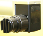
- In this procedure anterior most layer of cornea (surface layer or epithelium) is scratched / roughened in central 8 mm, and Riboflavin (vitB2) eye drops are instilled at every 5 min intervals for 25-30 minutes. Followed by UVA rays application on same area for next 30 minutes and applications of Riboflavin drops is continued.
- Eye is patched for 24-48 hours.
- As the beam is focused at 300 mm. It does not cause any damage to deep structure of eye.
- Oral painkillers are given for one or two days
Post Operative Care
- 4-5 days rest in recommended.
- Eye drops are given for 2-4 weeks.
- For first 48 hours is it advisable not to put any water in the eye as surface epithelium takes 48 hours to heal.
- After a month of operation if required final lens fitting can be done.
- 3 monthly follow up is advised to note progression.

Results
Collagen crosslinking has been found to stop the worsening in 90 – 95 % patient. It is also found to reduce the irregularity (astigmatism) of the cornea. It improved the best-corrected vision and improvement contact lens fitting. This procedure is being performed at our clinic since December 2007. Once 70 patients have been filled successfully with contact lenses. One of the patient required to undergo corneal transplant surgery.
Dry Eyes
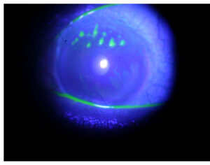
The eye depends on the flow of tears to provide constant moisture and lubrication to maintain vision and comfort. Tears are a combination of water, for moisture; oils, for lubrication; mucus, for even spreading; and antibodies and special proteins, for resistance to infection. Special glands located around the eye secrete these components. When there is an imbalance in this tear system, a person may experience dry eyes.
Symptoms:
Pain
Light sensitivity
A gritty sensation
A feeling of a foreign body or sand in the eye
Itching
Redness
Blurring of vision
Sometimes, excess tears running down the cheeks.
Causes:
An imbalance in the tear-flow system of the eye, causes situations that dry out the tear film. This can be due to dry air from air conditioning, heat, dust, cigarette smoke or other environmental conditions. Working long hours on the computer, the eye is constantly focused on the screen and neglects to blink enough to lubricate itself, causing an uncomfortable dryness. Long term contact lens usage can be a cause of dry eye syndrome – in fact, dry eyes are a rather common complaint amongst contact lens wearers. Other conditions that may cause dry eyes are:
- The natural aging process, especially menopause.
- Side effects of certain medications such as antihistamines and birth control pills.
- Diseases that affect the ability to make tears, such as Sjogren’s syndrome, rheumatoid arthritis, and collagen vascular diseases.
- Structural problems with the eye lids that don’t allow them to close properly.
Treatment:
Though dry eyes cannot be cured, there are a number of steps that can be taken to treat them. Treatments for dry eyes may include:
Artificial tear drops and ointments.
- It is the primary treatment for dry eye.
- If you have chronic dry eye, it is important to use the drops even when your eyes feel fine, to keep them lubricated.
- If your eyes dry out while you sleep, you can use a thicker lubricant, such as an ointment, at night.
Temporary punctal occlusion.
Sometimes it is necessary to close the ducts that drain tears out of the eye. This is first done via a painless test where a plug that will dissolve over a few days is inserted into the tear drain of the lower eyelid to determine whether permanent plugs can provide an adequate supply of tears.
Permanent punctal occlusion.
If temporary plugging of the tear drains works well, then silicone plugs (punctal occlusion) may be used. The plugs will hold tears around the eyes as long as they are in place. They can be removed.
Cyclosporine eye drops.
It is currently the only prescription eye drop that helps your eyes increase their own tear production with continued use.
Other medications.
Other medications, including topical steroids, may also be beneficial in some cases.
Surgery.
If needed, the ducts that drain tears into the nose can be permanently closed to allow more tears to remain around the eye. This is done with local anesthetic on an outpatient basis. There are no limitations in activity after having this surgery.
Special Precautions:
Rest your eyes: When reading, watching television or working at the computer, stop periodically – about once every 20 minutes – to give your eyes a break and remember to blink!
Protect your eyes: If you suspect that, your dry eye syndrome is being caused by the environment -then take steps to reduce your exposure to these elements.
a. Wear sunglasses whenever you head outdoors. Proper sunglasses, with adequate UV protection lenses and protective wraparound frames, not only shield your eyes from the sun, but also give an added layer of protection against wind, dirt and dust particles in the air.
b. At home, use an air cleaner to filter out dust and mould particles in the air.
c. If your home is air-conditioned or heated, get a humidifier to add moisture.
Avoid direct air-conditioning: Sit away from blasting air vents or turn the vents away from you. Heating or air-conditioning can dry out your eyes quickly.

Cataract
What is a Cataract?
Cataract is a clouding of the normally clear lens of the eye. The amount and pattern of cloudiness within the lens may vary. If cloudiness is in the center of the lens vision is affected more & if the cloudiness is in periphery of lens, you may not aware that a cataract is present.

What are the Common Symptoms?
- Painless blurring of vision
- Glare or light sensitivity
- Poor night vision
- Needing bright light to read
- Fading or yellowing of colors
What causes Cataract?
- Aging of the eye
- Injury to the eye
- Medical problem such as diabetes
- Medication, especially steroids
- Unprotected expose to Sunlight
- Previous eye surgery
How fast does a cataract develop?
It varies among individual and may even be different between two eyes. Most age related cataracts progress gradually over a period of year. Cataracts in patients with diabetes younger people may progress rapidly over a short time.
How is the Cataract detected?
A thorough eye examination by ophthalmologist (eye surgeon) can detect the presence of a cataract It will also rule out any other condition causing blurred vision or other eye problems and problem with other part of eye (such as the cornea, retina or optic nerve) that can affect visual improvement after cataract surgery. After a thorough eye examination ophthalmologist can tell you how much visual improvement is likely after surgery.
How is the Cataract treated?
Surgery is the only way a cataract can be removed. However if symptom of cataract are not bothering you very much, surgery may not be needed. No medication ,dietary supplement or exercise have been shown to prevent or cure cataracts Protection from excessive sunlight may helps to slow the progression of cataracts.
When should surgery be done?
Surgery should be considered when cataracts cause enough dimness of vision to interfere with your daily activities. It is not true that cataract need to be ‘ripe’ before it can be removed. You must decide if you can see well enough to do your job, drive safely and read or watch T V in comfort. Does your vision allow you to perform daily tasks like cooking, shopping, taking medication without difficulty? Based on the symptoms you and your ophthalmologist should decide together when surgery is appropriate.
What can I expect from cataract Surgery?
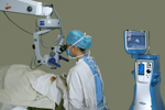
Cataract surgery is usually done under topical / local anesthesia as out patient procedure. During surgery cloudy lens is removed and it is replaced by a permanent intra ocular lens implant. After surgery you will have to take eye drops as directed by your ophthalmologist. After cataract surgery 95% of cases notice improvement in vision unless there is problem with other parts of eye. It is important to understand that complication can occur during or after the surgery, some severe enough to limit vision. If you experience slightest problem after surgery, contact your doctor immediately In many people who have cataract surgery the natural capsule that support the Intra Ocular lens become cloudy. It can be cleaned by laser procedure to restore vision.
POST OPERATIVE INSTRUCTIONS:
Take your normal diet or as permitted if you have diabetes or hypertension.
Take bath everyday , you can wash your hair avoiding the entry of water in the operated eye first week after operation.
Watch T.V. , read and write.
Climb or get down steps.
Take your medications like anti hypertensive or anti diabetic drugs according to your physician’s advice.
Wear your old glasses or wear dark glasses during daytime and wear the green plastic shield while sleeping ( day or night ) for 4 weeks.
KEEP A NOTE OF THE FOLLOWING :
Do not sleep on the side of your operated eye for two weeks.
Avoid lifting heavy weights.
Be careful with small children as they may hurt your eye unknowingly.
Do not drive car , two wheeler for three weeks.
Do not discontinue the medicines prescribed by us without our knowledge.
Avoid doing Namaz for one week.
GENERAL INSTRUCTIONS:
The person who is cleaning the eye or putting the eye drops should wash hands thoroughly with soap and water before putting the eye drops.
While putting the eye drops , pull the lower eyelid gently and instill one drop of prescribed medicine in the eye.
Replace the cap of the dropper immediately after use . do not let the dropper tip touch any surface.
Avoid putting the pressure on the eye.
Avoid putting the eye drops yourself and if you are not sure, ask your attendant to do it for you.
Clean the eye twice daily with sterilized cotton, if necessary.
Sterilize the cotton by boiling in water for 15 minutes and use when it is lukewarm.
Squeeze the water , ask the patient to close the eye , wipe gently from the corner outwards along the lashes. Change the cotton swab after use.
IF YOU EXPERIENCE ANY OF THE FOLLOWING SYMPTOMS , WHICH LAST MORE THAN 24 HOURS , NOTIFY OUR CLINIC OR AN OPHTHALMOLOGIST IMMEDIATELY . IT IS IMPORTANT THAT YOU BE AWARE OF THESE SYMPTOMS.
- ANY SUDDEN CHANGE IN VISION OR IN BLURRING OF VISION.
- INCREASE IN SENSITIVITY TO LIGHT , INCREASE IN REDNESS.
- A WHITE OR CLOUDY CORNEA OR PUPIL .
- PERSISTENT DISCOMFORT OR PAIN . IF THE PAIN IS EXTREMELY SEVERE , DO NOT WAIT FOR 24 HOURS.
Corneal transplant ( Keratoplasty)
Corneal transplantation / corneal Grafting / Keratoplasty is a surgery performed to replace diseased or affected part of the cornea.
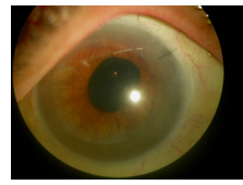
The cornea is the front surface of the eye. In health it is optically clear and allows a clear image to be formed on the retina. Conditions like infection, injury , dystrophy and degeneration can make this layer cloudy, resulting in Corneal blindness. The cornea is like a sandwich , made of many layers. The disease in the cornea can affect one or more layers.
Replacing the corneal layers to the affected depth can ‘clear ‘ the opacity. Hence Corneal Transplantation may be Full thickness ( Penetrating Keratoplasty) or partial thickness (Lamellar Keratoplasty).
If the superficial layers are transplanted , it is termed as an Anterior Lamellar keratoplasty , while if the innermost layer is affected (the endothelium , with a thin sliver of stroma) and replaced , it is called an Endothelial Keratoplasty.
The advantages of lamellar grafts is the benefit of closed eye surgery, reducing the risks of bleeding , hemorrhage, infection and most of all rejection. Corneal Transplantation is the most successful organ transplant with a success rate of 85 to 90 percent.

Painful red eye in arthritis
Rheumatoid Arthritis, Ankylosing Spondylitis, Psoriatic Arthropathy, Wegener’s, Polyarterirtis Nodosa, Lupus etc. are various auto immune diseases, where the immune system also targets the ocular tissues and other organs like the lungs and kidneys , besides causing destruction of the joints due to inflammation of the cartilages and synovial tissues.
Autoimmune reactions and inflammation mediated via Interleukins and T cell mediated antigen antibody reactions. HLA B-27 is known to be associated with tendency to recurrence of the disease, as it presents the altered antigen to the T cells. The ocular collagen is often misrecognized as the ‘target’ organ and immune mediated lesions occur in the various structures of the eye.
Ocular involvement in immune mediated arthritis may preceded or follow the manifestation of the systemic disease. Hence a painful red eye in a patient may be the presenting feature of a systemic disease.
The eyes can be affected in many ways. Conjunctival congestion, Secondary Sjogren’s syndrome or dry eyes, peripheral ulcerative keratitis or Mooren’s ulcer, marginal keratitis, sclero keratitis, episcleritis, scleritis and iridocyclitis are known manifestations of eye disease in immune mediated arthritis. Sterile melting of the cornea is another sinister complication of rheumatoid eye disease and can occur quite unsuspectingly.
Dry Eyes Disease affects almost 50 % of patients of RA. It is more common in women The patient experiences irritation, foreign body sensation, mucoid discharge, redness and blurry vision. Examination of the eyes shows a reduced tear secretion, increased tear film break up time, staining of the cornea and conjunctiva. The pathogenesis of dry eye in RA (like other forms of Sjogren’s) involves immune destruction of the goblet cells and lacrimal glands. The main stay of treatment is tear substitutes, lubricating gels or ointments and punctual plugs. Topical cyclosporine 0.05 or 0.1 % is recommended to increase the tear secretion. Systemic steroids and immunosuppression, as part of the therapy adds value to controlling the disease.
Episcleritis is a painless congestion of the episcleral tissues. It blanches with the instillation of epinephrine. The patient himself may not notice the redness and is often pointed out by other observers. This responds well to topical NSAIDs and may rarely need local steroid drops to be instilled.
Scleritits on the other hand is a painful mauve or violaceous congestion of the sclera. The patient complains of pain that may wake him up from his sleep. The area of congestion is tender and may extend under the lids and is hence more extensive that the seen lesion. Necrotizing sclerites is an extremely painful avascular necrosis of the sclera. The affected tissues show thinning and may become staphylomatous. Scleromalacia perforans, on the other hand is a painless necrosis and sloughing off of the sclera tissues. In all these conditions the vision remains unaffected unless it is accompanied by iridocyclitis or posterior uveitis. Posterior uveitis is often missed as the loss of vision is painless. Retinal detachment, sub retinal exudates and retinal hemorrhages are noted. When accompanied by panuveitis, the eye may become phthisic.
Iridocyclitis, can be acute (sudden) or chronic. The patient complains of haziness of vision along with pain and redness. There may be accompanying headache due to the secondary rise in Intraocular pressure. The cornea shows keratic precipitates, edema , anterior chamber flare and cells. The pupil is miosed and there may be posterior synechiae suggestive of previous episodes of uveitis. Subcapsular cataract may follow recurrent uveitis.
Marginal keratitis affects the peripheral cornea just within the limbus. There is congestion around the affected limbus. The peripheral cornea shows melting and thinning and may even perforate (more often following the instillation of topical steroids without systemic immunosuppression).
Sclerokeratitis occurs when the peripheral limbus and adjacent sclera is involved. Focal or diffuse keratitis is also noted . Sometimes there may occur an asymptomatic thinning of the cornea all around the limbus resulting in a ‘Contact lens’ cornea.
Investigation of a patient with arthritis and a red eye include all causes of uveitis / arthritis and few secondary causes.
Ocular lesions may be the first manifestation of the major systemic disease. Hence any patient with unexplained redness, corneal melts, marginal keratitis or peripheral ulcerative keratitis or uveitis must be investigated for a systemic disease. Haemogram, RA factor, C Reactive Protein, cANCA for Wegener’s and other vasculitis causes , pANCA for nonvasculitic causes like Ankylosing Spondylitis , ACE I level for Sarcoidosis, ANA and DsDNA for Lupus related arthritis. Dermatological opinion for psoriatic arthroathy , form the battery of tests that need to be done. An X-ray of the lumbosacral joint to rule out sacroiliitis and/or ankylosing spondylosis. Renal and liver function tests must be done before starting the immune suppressive agents as these are known to be hepato and nephrotoxic.
Treatment includes systemic steroids, Immunosuppressive, oral analgesics, topical steroids, NSAIDs, cyclosporine and cycloplegics. Additional therapeutic measures like tissue adhesives, patch grafts may be necessary.
Systemic steroids and Immunosuppression are essential in controlling the disease and so it’s ocular manifestations. After detailed investigations oral prednisolone is started at the dose of 1 mg/ kg to be tapered over two to three weeks. A low dose maintenance dose may be continued, ensuring the blood sugars as normal and calcium supplements given. Oral Methotrexate weekly 7.5 to 15 mg, or Azathioprine 100 mg daily. Anti TNF alpha agents like eternacept, infliximab have been used with good results.

Systemic therapy needs to be continued on a long term basis and hence monitoring under the care of a physician to be sure the disease process is under control and at the same time no toxicity involving bone marrow depression, renal or liver dysfunction occurs.
Local therapy in ocular lesions like Episcleritis, scleritis, iridocyclitis and keratitis are all treated with steroid eye drops. Prednisolone acetate 1% penetrates the best and has to be administered under close supervision. The dosage has to be titrated according to the local response. Any associated corneal thinning or melting must be treated with application of tissue glue, bandage contact lenses or patch graft with preserved cornea or sclera. Scleral thinning would require sclera patch grafts in areas of extensive thinning. Anti glaucoma drops and oral medication are required to control raised intraocular pressure resulting from the accompanying inflammation or the steroid response. Cycloplegics and antibiotics must also be prescribed. Artificial tears, lubricants and prophylactic antibiotic drops are adjuvants in the treatment.
Preservation of the ocular morbidity, by treating the active disease and preventing recurrences and preserving the vision and so the quality of life of the patient form the main aim of therapy of patients with ocular involvement in Arthritis.
Lasik and other refractive procedures
Refractive procedures are performed to reduce or remove the need for spectacles or contact lenses. It is best done after the refractive error stabilizes, ie., at least for two years, after the age of 18 years. Various procedures include, LASIK, SMILE, Advanced Surface Ablation, Intraocular Collamer Lens and Refractive Lens Exchange.
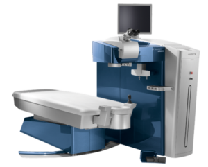

The choice of the procedure depends on, the amount of the refractive error, the corneal thickness and curvature and age of the patient.
LASIK
Is a procedure, where a flap of the cornea is made and raised. The laser beam is aligned on the cornea and the treatment applied, as per the pre-calculated data. The flap is replaced and eye drops instilled. The other eye is treated in a similar manner and the patient is made to wear a pair of dark glasses. Eye drops will be prescribed. A final check up will be done before you leave the center. Your appointment will be scheduled for the next day, with your doctor.
Femto Lasik, is the laser applied to make a flap. Microkeratome assisted – flap is made with an automated microkeratome.
Customized LASIK, includes treating aberrations, other than those corrected by spectacles or lenses.
The error that can be corrected is between -1.00 to -8.00 sph and +1.00 till +6.0 D sph. Astigmatism up to 4 D can be corrected.
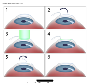
LASIK is a painless procedure. You may feel some discomfort for 4 to 6 hours after the procedure. You will be given some oral medication for the discomfort and you would best sleep out the discomfort. 95% of the vision is restores in 24 hours and the rest by 1 week. One can resume most activities in 24 to 48 hours.
SMILE
Small Incision Lenticule Extraction: is a new technology, which alters the shape of the cornea, by removing a disc of corneal tissue through a small incision. The advantage is that there is a small tunnel and no flap. Chances of dryness or injury to the flap are averted and the strength is maintained. However some restrictions remain pertaining to non correction of astigmatism over 2 D. Enhancement or retouch up if needed at a late date may be difficult.
ADVANCED SURFACE ABLATION
This technique is offered to patients as an alternative to LASIK, for lower errors ie., less than 4 or where the error is more than -8.0 or if the corneal thickness is on the borderline for LASIK.
- It is predictable, but there is a small chance of some error relapsing.
- Vision takes time to stabilize ie., upto 8 to 12 weeks.
- Slightly more painful, than LASIK.
- But offers a hope to patients with high errors where SMILE and LASIK cannot be offered.
ICL: INTRAOCULAR COLLAMER LENS
For patients whose cornea is too thin to undergo Lasik, a soft foldable lens can be inserted to correct the refractive error to as close to zero as possible. This is suitable for patients between the age of 18 to 35 years. It is a safe procedure.
SUPRACOR
For near vision correction after the age of 40 years supracor treatment is offered. This will reduce the dependence of glasses.
Diabetes and your eyes
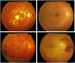
Diabetes affects the blood vessels of the entire body, but damage is mostly seen in the blood vessels of the eye (retina), kidneys, heart, and brain. Blood vessel damage leads to their blockage, bursting seen as bleeding, leakage of blood proteins and water, and formation of new blood vessels.
The retina is a light-sensitive layer located at the back of the eye. Each of these processes has a different effect on the delicate retinal structure. It is the only part of the human body where we can directly see blood vessels, and assess the effects of diseases like diabetes.
Diabetic Retinopathy (DR) is an important cause of permanent vision loss, and can even lead to blindness. DR proceeds through various stages. In the early stages of the disease, the patient most often does not have any vision-related complaints. If such a stage is reached, treatment can be initiated to preserve vision for the rest of one’s life.
Two main retinal problems are seen in DR – new blood vessel formation, and leakage in the macula (the central part of the retina). These conditions require different treatments.
In general, DR can be treated by retinal laser, injections, surgery, or a combination of these. Treatment, when warranted, is safe and effective.
Prevention is the best strategy – a combination of Regular Retinal Examination (advised by your retina specialist), and Good Diabetic Control are the keys to vision preservation.
Dr. Devendra Venkatramani is a proud alumnus of the prestigious Sankara Nethralaya Eye Hospital, Chennai. He underwent rigorous training in the medical and surgical management of retinal diseases at LV Prasad Eye Institute, Hyderabad. He has been awarded the coveted FRCS and FICO degrees.
Dr. Devendra is a consultant vitreo-retinal surgeon at Ruby Hall Clinic, Pune. He is also available at the Wagh Eye clinic , Ghole Road for consultation by prior appointment on Saturdays
Eye donation
1. What is Eye Donation?
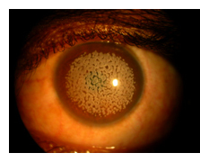
Eye donation means donating one’s eyes after death. The donated eyes are stored in a specialized laboratory called as Eye Bank. This is similar to a blood bank, storing tissues till the time there is a requirement of the eyes (cornea) for transplantation.
2. Who can donate eyes?
Anyone can donate eyes; there is no age bar for donation, male or female, spectacle wearers can also donate their eyes. Patients with hypertension, diabetics, asthmatics and those who have undergone surgery in the past whether of eyes or any other site on the body can also pledge to donate eyes.
3. Who cannot donate eyes?
Persons suffering from HIV/AIDS, Hepatitis B & C, Blood cancers, Rabies, Septicemia (infection throughout the body) cannot be considered for eye donation.
4. How can one donate eyes?
During life one can register his/her name with the closest eye bank by filling up pledge cards or after death the family members can request for an eye donation to the closest eye bank. Either of the cases the 1st blood related family member of the deceased has to give consent for eye donation. One should contact the eye bank within 6hrs of death.
5. Who can benefit from eye donation?
Cornea is the outermost, thin and transparent tissue of the eye through which light passes inside and we are able to see. Thereby only those people who are blinded because of damage to the cornea can be benefited from eye donation / eye (corneal) transplantation. Thus in corneal transplantation (keratoplasty) only the layer of cornea is replaced and not the entire eye as it is usually understood.
6. What are the causes of corneal blindness?
The majority of causes of corneal blindness are preventable. The common causes are infections, trauma (injury), chemical burns and some hereditary abnormalities. All the above mentioned conditions need transplantation when cornea loses its clarity.
7. What are the instructions for eye donation?
One should call the nearest eye bank within 6hrs of the death, give the proper address and telephone number of the location. By the time the eye bank team arrives:- • Close the eyelids • Place wet cotton /cloth over the closed eye lids • Elevate the head with pillows under the neck • Switch off the fans • If A/C is available switch it on
8. Why donate eyes?
In India: Number of corneal blind persons ——- 22 Lac Corneal Requirement every year ——– 75,000 to 1lac Total corneas collected every year——-33,000 Percentage of corneal blindness ———60% (under the age of 12yrs) 90% (under the age of 45 yrs) It’s obviously very clear that a great disproportionate ratio exists between demand and supply of corneal tissues and thereby it’s our moral duty to return to the society and the needy few the gift of sight.
9. What can YOU do for Eye donation?
One should register his/her name and pledge for eye donation with any eye bank. The most important action you can take to ensure you will be a donor is to tell your family and legal representative. The eye banking law requires your “Next of Kin” to give consent and medical history for the donation. Various NGO’s should take the initiative to promote eye donation campaigns in the society and organize meetings to create awareness and clarify the myths with eye donation.
10. What are the myths and facts of eye donation?
Myths Vs Facts
- My religion doesn’t allow organ and tissue donation. Most religions throughout the world support organ and tissue donation as a humanitarian act.
- If I donate my eyes it will disfigure my face As only the cornea is removed there is no disfigurement
- Donation will be costly to my family There is no cost to the donor’s family. The Eye Bank pays for all costs related to corneal donation
- The whole eye can be transplanted Only the cornea and the sclera (the white of the eye) can be transplanted
- Is cancer a rule-out for donation? No, not all cancers are contra-indications for eye donation.
- Eyes which have undergone surgery cannot be used Even eyes which have undergone surgery in the past can be used for transplantation.
Recent advances in cataract surgery
From days of Sushruta where cataracts were couched with red hot needle (practically a stitch-less , even smaller than keyhole ) we have come back to the era of “ suture-less” tunneled surgery. In the past cataract surgery with traditional sutures( stitches) and lenses needed the patient to be lying still in bed for a week, one month of rigorous restrictions and the outcomes for the vision were not predictable. The use of an operating microscope techniques to dissolve the lens and insertion of injectable lenses has revolutionized cataract surgery to an outpatient day care procedure , where the patient can resume work in less than a week.
The newer improvisation aim at emmetropic or perfect uncorrected vision. This does away with the need of any visual aids (eg. Spectacles). Phacoemulsification technique has become the gold standard of cataract surgery, where the cataract is emulsified with ultrasonic energy and then aspirated or sucked out with high vacuum systems, though self sealing valved incisions. This closed eye surgery prevents any leakage or ingress of infections or bleeding, hence minimizing risks. Routine phacoemulsification is performed through 2.2 mm tunnel. Further advances include sub 0.9 mm incision (or the microphaco or MICS-minimally invasive cataract surgery).It also enables patients to get back to their routine , almost immediately.
The so called ‘LASER CATARACT ‘ surgery is performed by using a ‘ femtosecond laser’ system to perform cataract surgery. This laser ensures very precise cuts without damaging surrounding structures and reduces the risk to the minimum.
As important as the technique of removing the cataract ,is the lens that is implanted during surgery. Intraocular lenses of appropriate power (calculated with sophisticated instruments)are inserted in place of the original lens to restore the sight.
Emmetropia or achieving 6/6 visual acuity is the aim of a perfect surgery. Intraocular lenses can be rigid (inserted through larger incision), foldable (injectable)injected through 2.2 or 0.9 mm tunnel incisions. The materials are polymers with UV blocking properties. Toric lenses neutralize astigmatism ensuring sharp and clear vision for distance or near. Monofocal lenses IOLs correct the power either for distance or near while multifocal lenses and accommodative ensure sharp focused vision for near, distance and intermediate vision.
Research edges towards the development of a ‘ no touch’ technique and restoration of perfect and crisp vision. However the technological sophistication comes at a price. The escalating costs of surgery are due to the expensive and technologically advanced equipment that the surgery demands .
Cataract blindness is the second most common cause for visual deprivation. Techniques like small incision cataract surgery , which use minimum instrumentation and indigenously produced intraocular implants aim to make this surgery affordable . NGOs and Government sponsored National programs , also ensure that cataract surgery is within the reach of one and all.